■Direct observation of the epidermis
Even when you observe the hair at high magnification it is difficult to see the cuticle (The black foreign object simply becomes larger.)
Observe with ring light
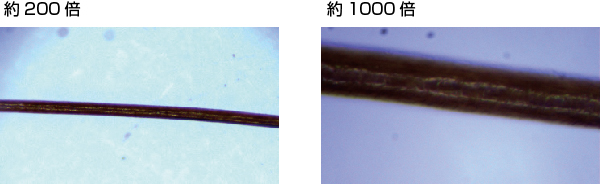
If it’s a “high magnification type with coaxial illumination” it’s more or less easier to see.
Furthermore, the resolution of the lens must also be high.
(1) Universal high magnification microscope (coaxial illumination)
NSH130CS-R ultra-high magnification microscope, but low lens resolution (coaxial illumination)
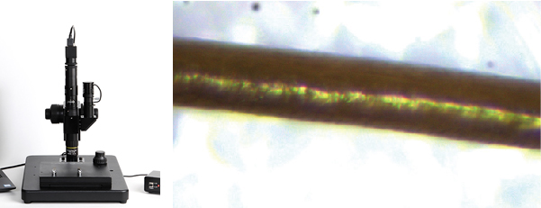
Due to the low resolution of the lens, the outline of the epidermis is blurred and cannot be observed.
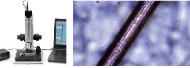
(2) High resolution/high magnification microscope (coaxial illumination)
Ultra high magnification and high resolution USB CCD microscope USH130CS-H1 Ultra high magnification microscope with high lens resolution (coaxial illumination)
(3) Metallurgical microscope
GR3400J Metallurgical Microscope Metallurgical microscope is a coaxial illumination microscope. Although not intended for use, you can clearly see the cuticles. If you just want to look at the cuticles, I think that’s the most cost-effective method.
To actually take pictures like the one below, you’ll need an electron microscope.*We don’t handle electron microscopes.
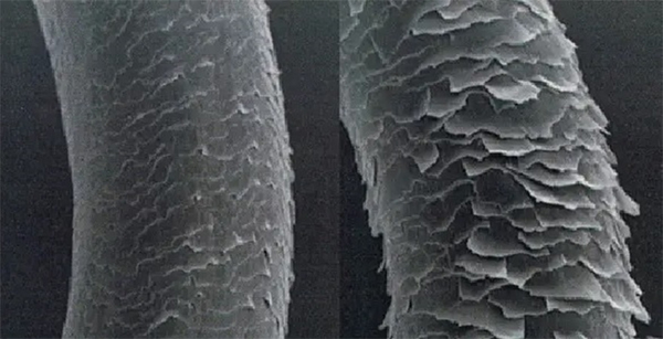
<Comparative image of epidermal observation>
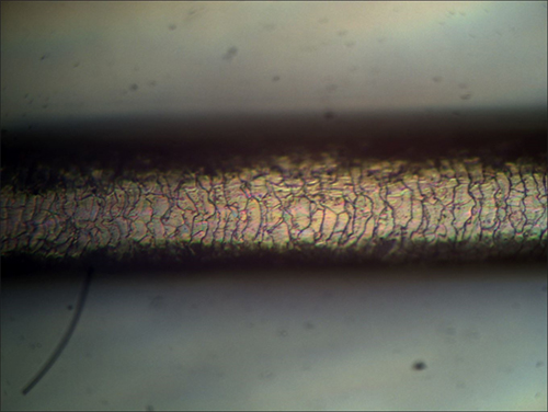
■Metal microscope + 400x microscope camera
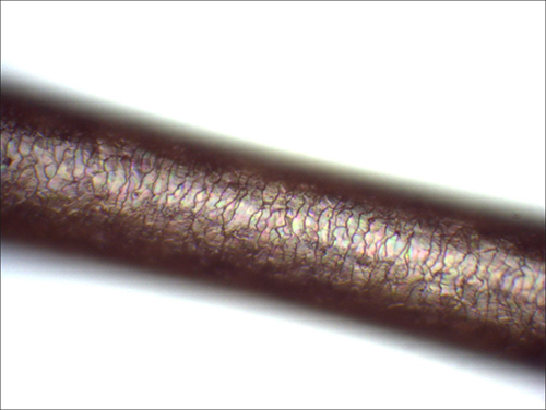
■Ultra high magnification microscope (USH3130CS-H1) 800x
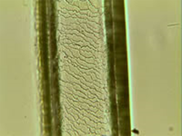
■Indirect observation of the epidermis
The epidermis cannot be directly observed using a biological microscope.
However, it can be observed indirectly using a technique called the reservoir method
Prepare the “sewage liquid” and “sewage plate”.

Place the hair on the sump, apply the liquid in the tank and let it sit for a while.
The surface of the hair is transferred to the wastewater plate.
Observe this table using a biological microscope.
The following is an image of the epidermis observed at our company using the Sumpu method.